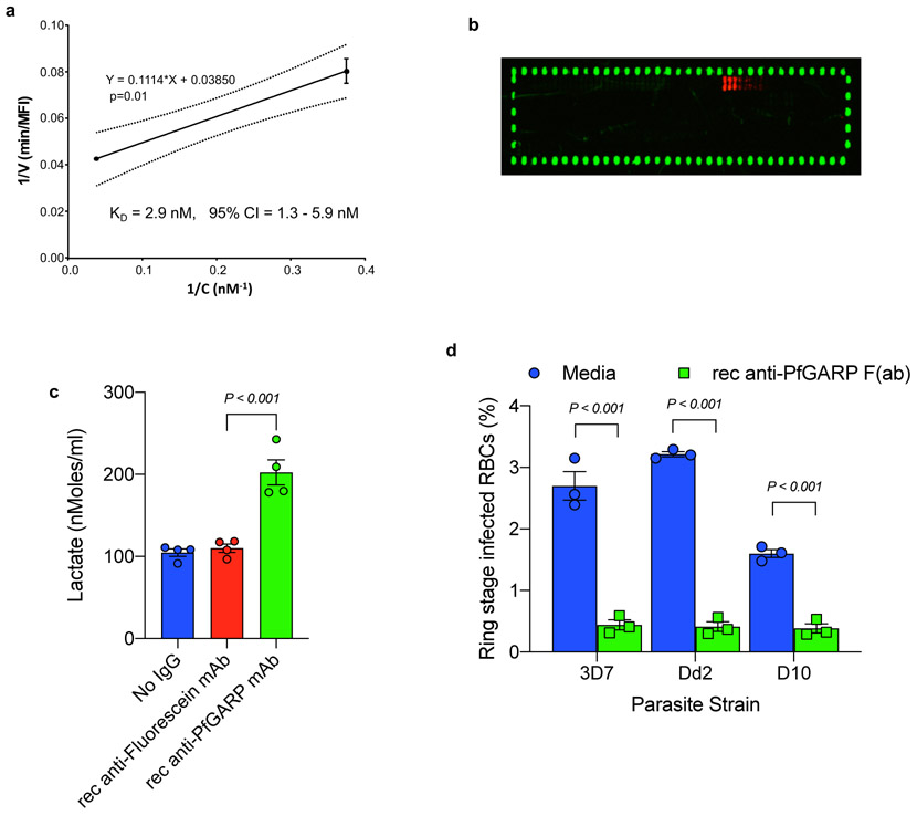
Extended Data Figure 2 ∣. Characterization of recombinant monoclonal anti-PfGARP.
a) Kinetics of binding between rec mAb7899 and PfGARP-A was measured at two antibody concentrations. For each concentration, two biologically independent replicates were performed. The experiment was performed twice and the data was pooled. The 95% confidence intervals (CI) is shown by dashed lines. Error bars represent standard. Formula shows linear regression. V, initial velocity of binding; c, concentration of biotinylated rec mAb7899 ; min, minutes, MFI, Median Fluorescence Intensity. b) Epitope mapping of rec mAb7899. We printed a custom 15-mer peptide microarray contained 264 different peptides which spanned the PfGARP-A sequence (aa 410-673). The peptides overlapped by a single amino acid and were printed in duplicate, framed by HA control peptides. The array was probed with rec mAb7899 (red) and anti-HA (green) and imaged on a LI-COR Odyssey. c) Ring stage 3D7 parasites were cultured in the presence of media, recombinant mAb anti-PfGARP, or recombinant mAb anti-fluorescein. Parasites were cultured for 48 hours at 37°C and lactate levels were measured in culture supernatant. Bars represent means of 4 biologically independent replicates, error bars represent SEMs. P value calculated by non-parametric two-sided Mann-Whitney U test is indicated. d) Ring stage 3D7, D10, or Dd2 parasites were cultured in the presence of media alone or recombinant anti-PfGARP Fab antibody (1 mg/ml) for 48 hrs at 37°C and ring or early trophozoite stage parasites were enumerated by microscopy. Bars represent means of 3 biologically independent replicates, error bars represent SEMs. P values calculated by non-parametric two-sided Mann-Whitney U test are indicated. Data representative of two biologically independent experiments.

