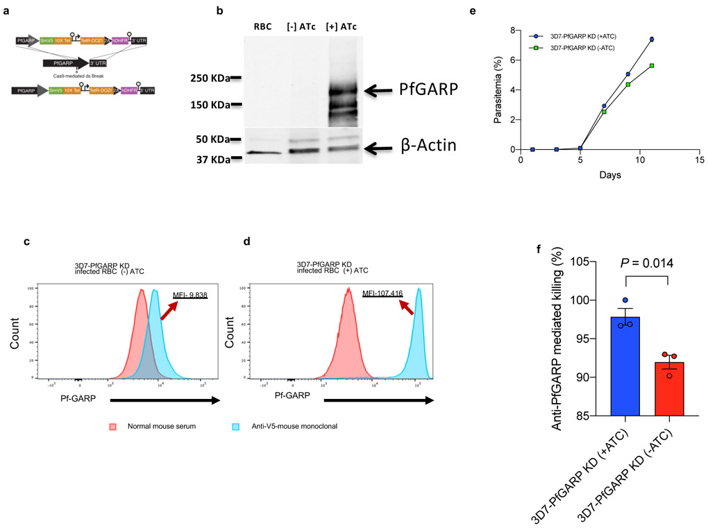
Extended Data Figure 3 ∣. Construction and characterization of PfGARP knock down parasite line.
a) Targeting strategy for creating 3D7-PfGARP KD parasites, b) Immunoblot analysis of 3D7-PfGARP KD. Parasites were sorbitol-synchronized at the ring stage and incubated with or without anhydrotetracycline (Atc) for 20 hours and probed with anti-V5. The expected molecular weight of the PfGARP-V5 tagged protein is 124 kDa, however its apparent mobility is 165 kDa due to its acidic composition. Lane 1, uninfected RBCs, Lane 2- 3D7-PfGARP KD infected RBCs cultured without ATc, lane 3, 3D7-PfGARP KD infected RBCs cultured with ATc. c and d) Expression of PfGARP on the surface of human RBCs infected with 3D7-PfGARP KD parasites. Ring stage 3D7-PfGARP KD parasites were cultured to the trophozoite stage in the absence (C) or presence (D) of ATc. PfGARP expression on surface of infected human RBCs (fixed but not permeabilized) was determined by flow cytometry, using monoclonal anti-V5 as primary and anti-mouse IgG-FITC as secondary antibody. Infected RBCs were gated and identified as described in Fig. 1e. e) Growth curves for 3D7-PfGARP KD parasites. Ring-stage parasites were cultured with or without ATc. Parasitemia was measured by microscopy. Each data point represents the mean of 3 biologically independent replicates, error bars indicate SEM. f) Growth inhibition assay using 10% anti-rPfGARP-A antisera or pre-immune antisera on 3D7-PfGARP-KD parasites cultured with or without ATC. Bars represent means of 3 biologically independent replicates, error bars represent SEM. P value calculated by non-parametric two-sided Mann-Whitney U test are indicated. Panels b, d, e, and f are representative of 3 independent experiments. Panel c is representative of 5 independent experiments.

