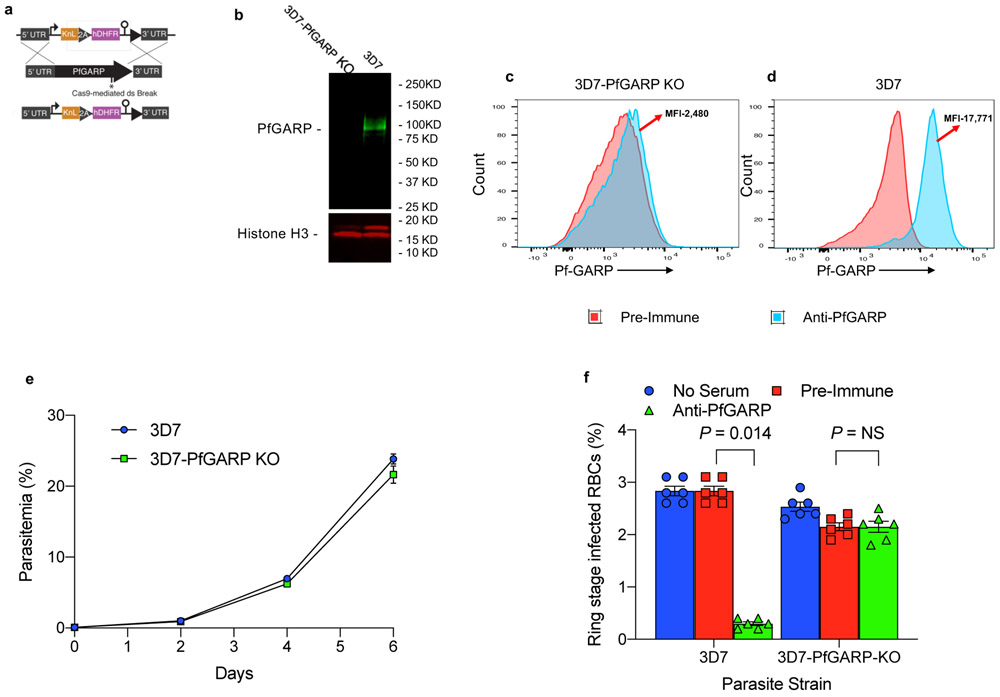
Extended Data Figure 4 ∣. Construction and characterization of PfGARP knock out parasite line.
a) Targeting strategy for creating PfGARP KO parasites, b) Immunoblot analysis of 3D7-PfGARP-KO. Trophozoite stage 3D7 or 3D7-PfGARP KO parasites were probed with anti-PfGARP and anti-actin as a loading control. Lane 1, 3D7-PfGARP KO infected RBCs, Lane 2- 3D7 infected RBCs. c and d) Expression of PfGARP on the surface of human RBCs infected with 3D7-PfGARP KO parasites. Ring-stage stage 3D7 or 3D7-PfGARP KO parasites were cultured to the trophozoite. PfGARP expression on the surface of infected human RBCs (fixed but not permeabilized) was determined by flow cytometry, using anti-PfGARP as primary and anti-mouse IgG-FITC as secondary antibody. Infected RBCs were gated and identified as described in Fig. 1e. e) Growth curve for 3D7-PfGARP KO parasites. Ring-stage stage 3D7 or 3D7-PfGARP KO parasites were plated at 0.5% parasitemia and cultured for 6 days. Parasitemia was measured by microscopy. Each data point represents the mean of 3 biologically independent replicates, error bars indicate SEM. f) Ring stage 3D7 or 3D7-PfGARP KO parasites with targeted deletion of PfGARP were cultured at 1:10 dilution in the presence of anti-PfGARP-A mouse sera generated by mRNA LNP immunization. Negative controls included no anti-sera and pre-immune mouse sera. Parasites were cultured for 48 hours at 37°C and ring and early trophozoite stage parasites were enumerated by microscopy. Bars represent the mean of 6 biologically independent replicates, error bars represent SEMs. P values calculated by non-parametric two-tailed Mann-Whitney U test are indicated. Data are representative of two independent experiments.

