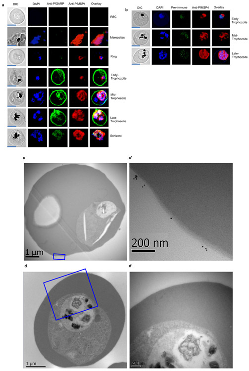
Extended Data Figure 5 ∣. Immunolocalization of PfGARP.
a) Uninfected and infected RBCs were probed with mouse anti-PfGARP prepared by DNA vaccination (green) and rabbit anti–PfMSP-4 (red) and counterstained with 4′,6′-diamidino-2-phenylindole (DAPI) to label parasite nuclei. PfGARP is detected on early, mid and late-trophozoite-infected RBC membranes and does not co-localize with PfMSP-4 (which localizes to the parasite membrane, scale bars 5 μm). b) Trophozoite-infected RBCs do not label when probed with pre-immune mouse sera by immunofluorescence microscopy (scale bars 5 μm). Non-permeabilized, un-fixed trophozoite infected RBCs (c and d) were incubated with anti-rPfGARP polyclonal (c) or control mouse serum (d), probed with anti-mouse IgG labeled with 10 nm gold particles, fixed, embedded and visualized by transmission electron microscopy. PfGARP localized to the outer leaflet of trophozoite infected RBCs. Figures labeled with primes represent higher magnifications views of the parent figure. Panel a and b are representative of 5 biologically independent experiments. Panel c and d are representative of 2 biologically independent experiments.

