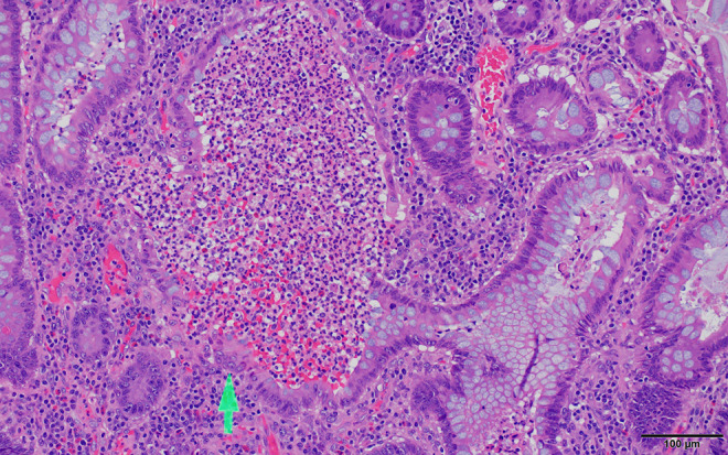
Figure 6.

Ulcerative colitis biopsy histology. Dilated glands containing inflammatory cells which at high magnification are recognized as neutrophils, forming crypt abscesses (arrow) typical of ulcerative colitis (UC) but which can also be seen in Crohn’s disease (CD). Scale bar: 100 µm.
