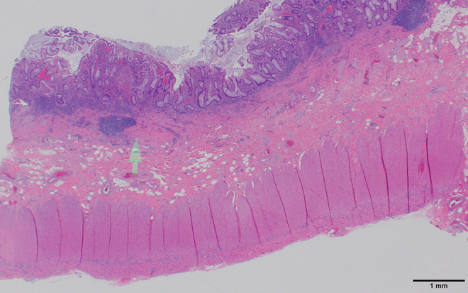
Figure 8.

Ulcerative colitis histology. The low-power view shows a markedly expanded lamina propria inflammatory cell component which does not extend into the submucosa (arrow). There is crypt distortion with irregular shaped and branched glands as well as loss of glands which are features of chronicity (also seen in Crohn’s disease) separating inflammatory bowel disease from acute self-limited (infectious) colitis. Scale bar: 1 mm.
