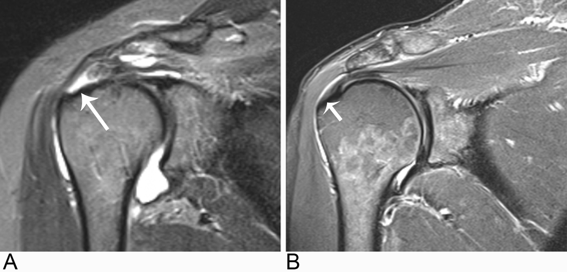
Fig. 1 –

Shoulder MR image examples of study participants in each cohort.
A, Oblique coronal STIR MR image from a study participant in the painful full-thickness supraspinatus tendon (SST) tear (long arrow) cohort.
B, Oblique coronal STIR MR image demonstrating no full-thickness SST tear (short arrow) in a study participant from the control cohort.
