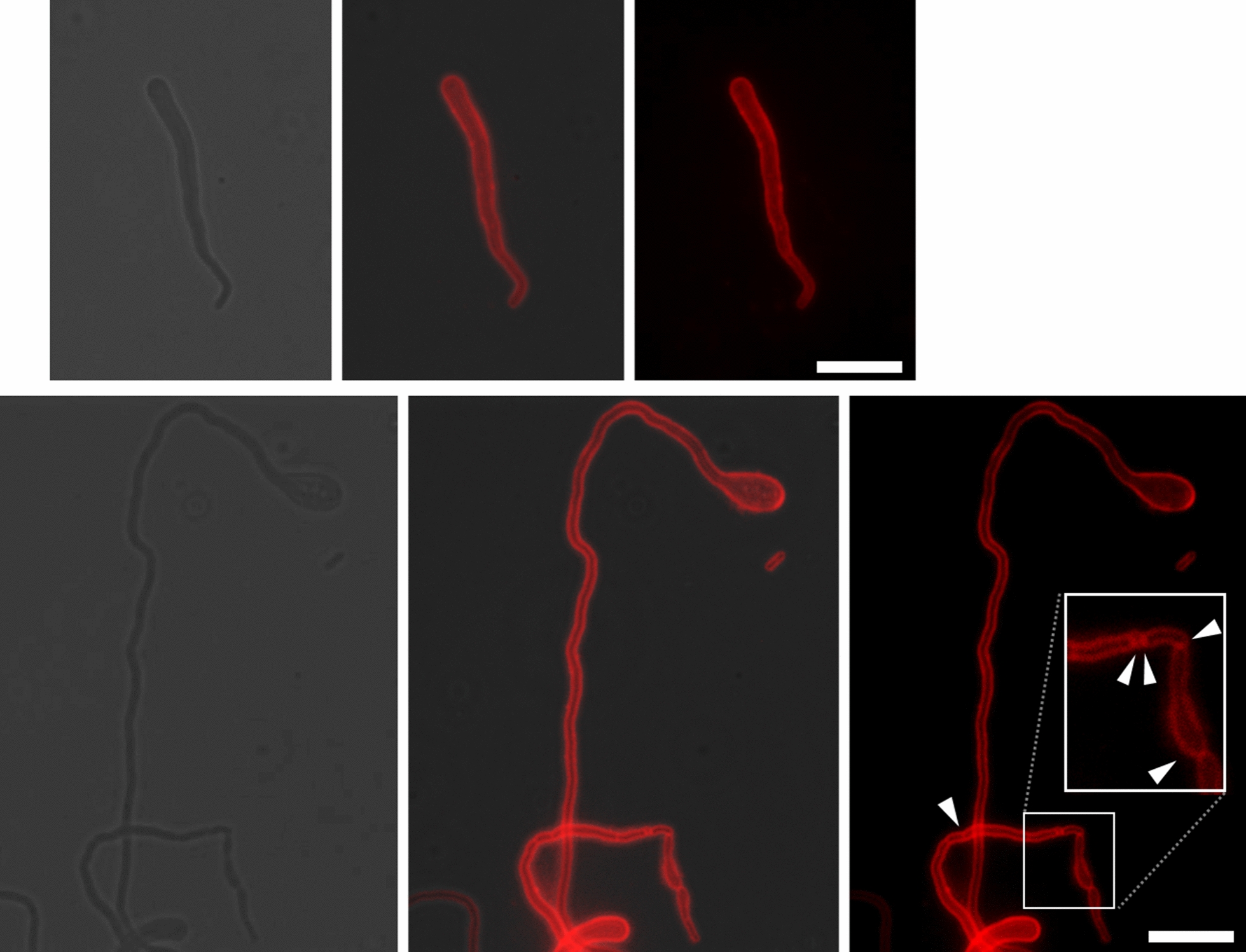
Fig. 6.

The membrane staining of Zm6 and KFS1 strains growing under the salt condition. The growing Zm6 and KFS1 cells under salt condition (NaCl 0.225 M) was stained by Fm4-64 at concentration of 20 μg.ml−1 for 15 min for the membrane visualization. The dye was washed by PBS prior to mounting on the agarose-pad for fluorescent microscopy. Imaging revealed that the bulged Zm6 cell (top panels) did not show any septa nor membrane compartment inside of cells. The KFS1 cell (bottom panels) occasionally formed very long filaments over 50 μm without any septa within cell. Locally frequent septa formation was found in some KFS1 cells, as pointed by white arrowheads. These suggest that septation was not tightly controlled in the strain under the salt condition. Red; Fm4-64 fluorescent signal. Scale bar; 10 μm
