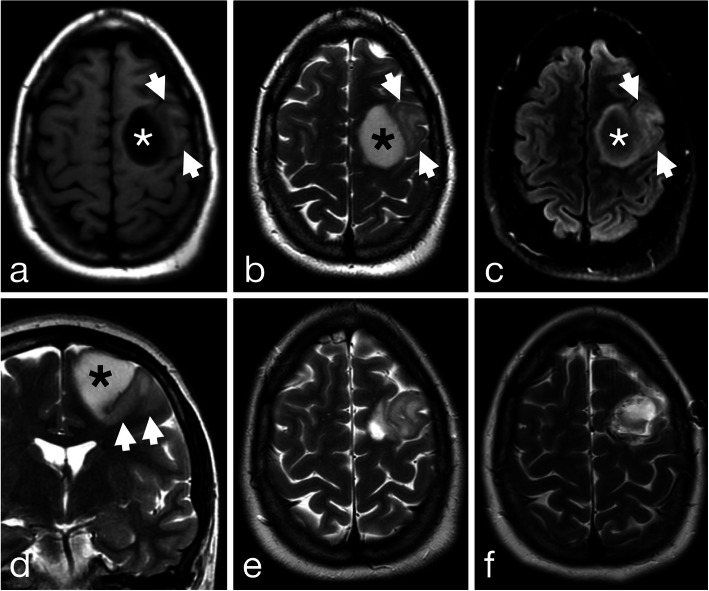
Fig. 1.
High left frontal tumor with two radiologically distinct components on preoperative axial T1-weighted (a), T2-weighted (b), FLAIR and coronal T2-weighted (d) MR images. A medial T1-hypointense T2-hyperintense component (asterisk) centered in the superior frontal gyrus shows well defined margins and the “T2-FLAIR” mismatch sign indicative of IDH1 mutant 1p/19q non-codeleted astrocytoma. A contiguous more lateral component (arrowheads) centered in the middle frontal gyrus shows less well-defined margins with cortical infiltration and gyral expansion more typical of oligodendroglioma. Follow-up axial T2-weighted imaging after resection of the more medial component (e) and the more lateral component (f)

