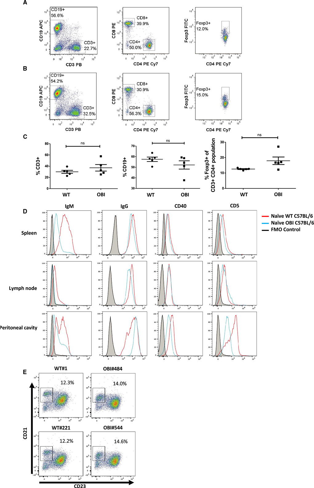
FIGURE 2.

B cell, CD4+ T cell, CD8+ T cell, and Treg compartments show similar proportions in (A) naïve WT C57BL/6 and (B) naïve OBI splenocytes. C, Analysis of spleen samples from multiple OBI and WT C57BL/6 mice. D, Naïve WT and OBI B cells express similar levels of IgG, CD40, and CD5 in spleen, LN, and PC. OBI B cells express a lower level of IgM compared to WT B cells in all 3 compartments. Spleen data represent 5 individual experiments. LN and PC data represent 3 individual experiments. E, CD19+ B cells isolated from the spleen from OBI and WT mice show present populations of marginal zone and follicular B cells. Data from 2 independent experiments are shown (left column: WT, right column: OBI). FITC, fluorescein isothiocyanate; LN, lymph node; OBI, ovalbumin (OVA)-specific BCR mice; PC, peritoneal cavity; Treg, regulatory T cell; WT, wild type
