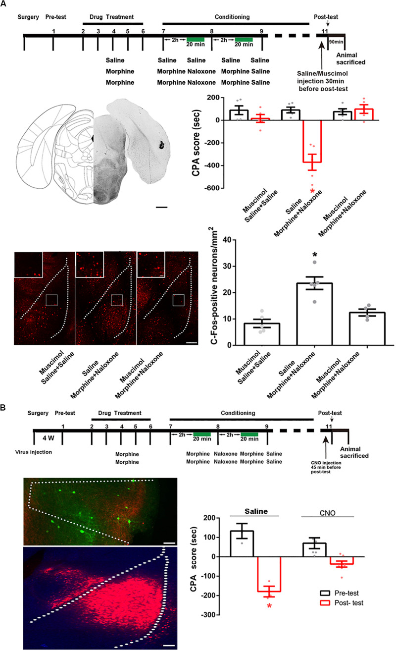
FIGURE 8.
The influence of inactivation of the POR on the increased expression of c-Fos in the BLA and the influence of chemical-genetic inactivation of the projection neurons from the POR to the BLA on CPA score in morphine withdrawn mice. (A) The influence of the inactivation of the POR on the increased expression of c-Fos in the BLA. Top panel: experimental timeline and groups for the CPA procedure. Left middle panel: the typical injection site of muscimol in the POR. Scale bar = 500 μm. Right middle panel: the CPA score of each group (n = 5 in each group, *p < 0.0001, compared with pre-test, two-way ANOVA, Bonferroni post hoc analysis). Left down panel: C-Fos positive neurons in the BLA in each group. Scale bar = 100 μm. Higher magnification images of boxed regions are shown on the top. Scale bar = 20 μm. Right down panel: the average numbers of c-Fos positive neurons in the BLA in each group (n = 5 in saline + saline + muscimol group and morphine + naloxone + saline group, n = 4 in morphine + naloxone + muscimol group, *p = 0.0003, one-way ANOVA following by Tukey post hoc analysis). (B) The influence of chemical-genetic inactivation of the projection neurons from the POR to the BLA on CPA score in morphine withdrawn mice. Top panel: the CNO inhibit experimental timeline and groups for the CPA procedure. Left middle panel: expression of hM4Di (Gi) (green-colored) in the POR 4 weeks after virus injection. Scale bar = 100 μm. Left down panel: expression of WGA-Cre (red-colored) in the BLA 4 weeks after virus injection. Scale bar = 100 μm. Right down panel: the CPA scores of saline group and CNO group (n = 7 in CNO group, n = 4 in saline group, *p = 0.0009, compared with pre-test, two-way ANOVA, Bonferroni post hoc analysis). Data are shown as the mean ± SEM.

