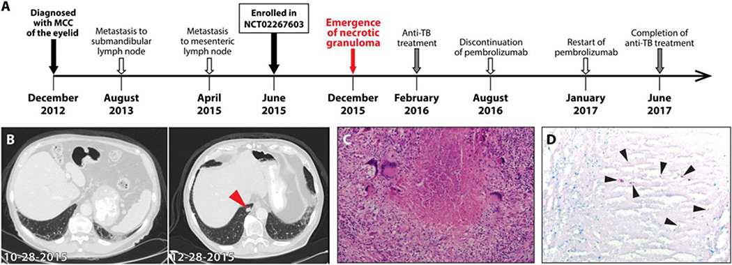
Figure 2. Development of tuberculosis in a patient treated with pembrolizumab for Merkel cell carcinoma.
(A) The timeline of therapy and disease status of a patient with Merkel cell carcinoma (MCC). (B) The chest CT images of a patient ~5 months after initiation of PD-1 blockade (10–28-2015, left) and ~2 months later (12–28-2015, right). Arrow head indicates necrotizing nodule in the right lower lobe. (C) The hematoxylin and eosin stain of the excised lung nodule with necrotic center surrounded by multinucleated giant cells. (D) The result of acid-fast stain of the lung nodule. Arrow heads denote AFB in the granuloma.

