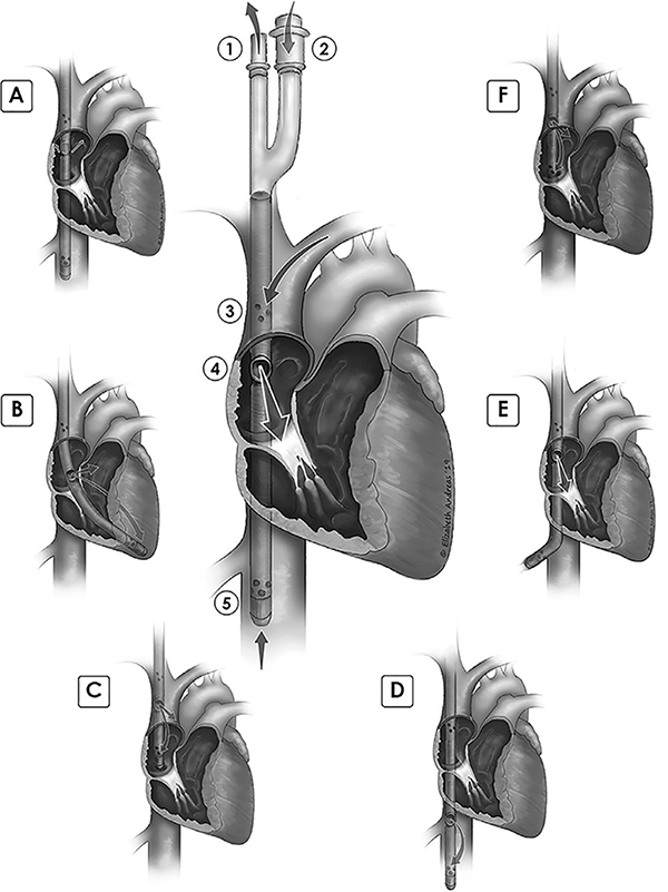
Figure 1: Cannula correct position and malposition illustrations.
Central figure: proper cannula depth and orientation. 1. Bicaval drainage outflow limb. 2. Post-oxygenator return limb. 3. Superior vena cava drainage orifices. 4. Return flow spout, at level of upper fossa ovalis, return jet directed anteriorly, towards center of tricuspid valve. 5. Inferior vena cava drainage orifices, with distal tip located a few centimeters beyond hepatic vein. Malposition illustrations and case numbers: A. Return flow oriented away from tricuspid, jet directed posteriorly, towards interatrial septum- 4 cases. B. Cannula within right ventricle, return flow recirculating by entering distal drainage orifices. Tip is against distal RV lateral wall- 2 cases. C. Distal tip of cannula in right atrium, too shallow. Return flow jet within SVC, likely not visible by TEE. Recirculation of flow from return jet into distal drain orifices- 6 cases. D. Cannula too deep, return flow within IVC, recirculation into distal drainage orifices- 7 cases. E. Tip in hepatic vein. Vein may collapse around distal drainage orifices, leading to chatter and low flow alarms in ECMO circuit- 6 cases. F. Return flow at upper RA/SVC junction, leading to recirculation of flow with some return flow entering distal orifices; distal tip is near RA/IVC junction; note that return flow may be seen with TEE- 3 cases.

