
Fig. 2.
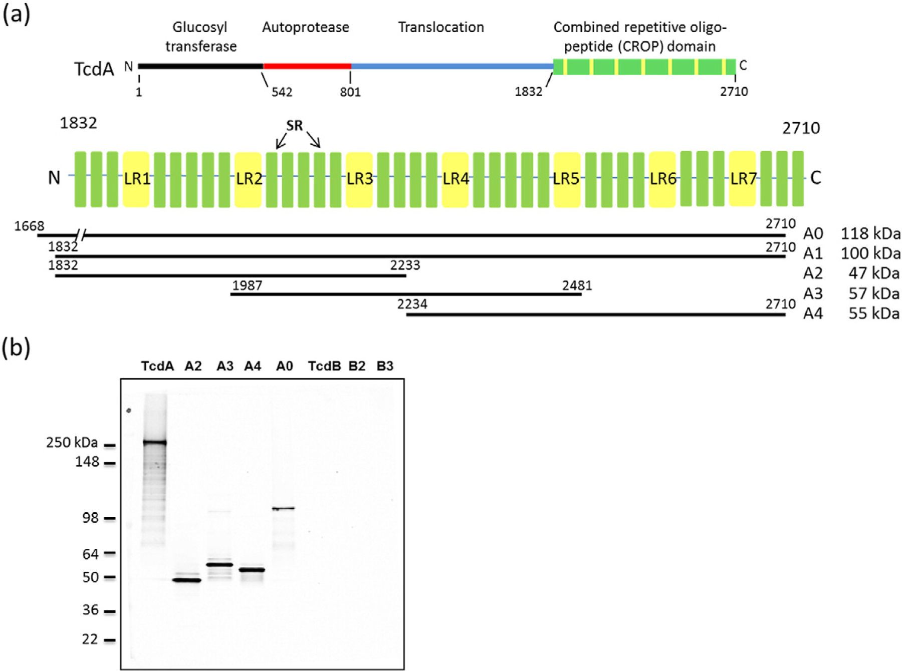
TcdA constructs used in this study and binding of actoxumab by Western blotting. (a) Domain organization of TcdA showing the CROP domain with LRs and SRs highlighted. The CROP fragment constructs (A0, A1, A2, A3, A4, and A5) used in this study are also shown. (b) Western blot of TcdA and TcdB holotoxins and of constructs A0, A2, A3, A4, B2, and B3.