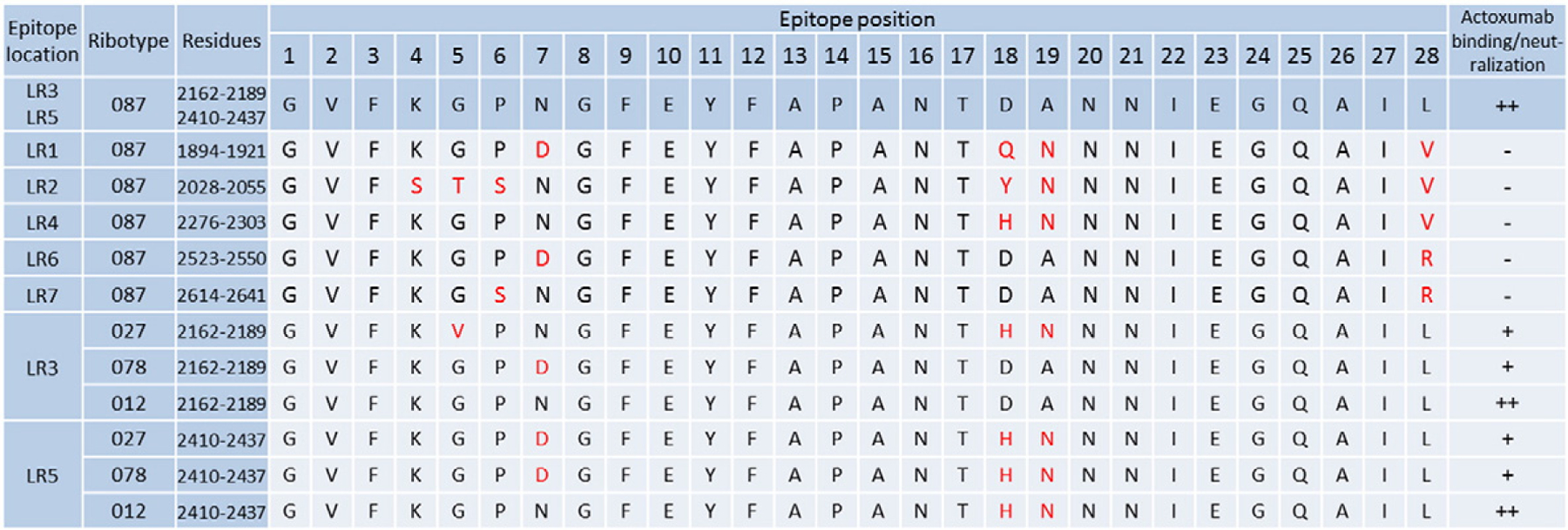
Fig. 6.

Alignment of actoxumab epitope sequences in TcdA. Residues within the putative actoxumab epitopes at LR3 and LR5 of ribotype 087 (VPI 10463) TcdA (as identified by HDX-MS) are shown by position (1–28) and compared to the homologous repeat sequences (LR1, LR2, LR4, LR6, and LR7) in the TcdA CROP domain and to the corresponding LR3 and LR5 epitopes within TcdA of ribotypes 027, 078, and 012. Residues that are different from those found at the same position in the actoxumab epitopes (LR 3 and 5) of ribotype 087 TcdA are marked in red. The ability of actoxumab to bind to individual homologous regions (as determined in this report) orto neutralize TcdA from various ribotypes (as demonstrated in Ref. [35]) is indicated (-, no binding; +, moderate neutralization; ++, high binding/neutralization).
