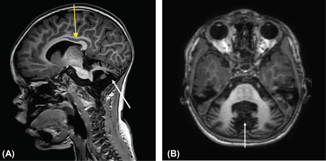
Figure 1. Twenty-one month old child with cerebellar hypoplasia.
(A) Mid-sagittal T1-weighted image showing thinning of corpus callosum (yellow arrow) and decreased cerebellar volume (white arrow). (B) Axial T1-weighted image showing decreased volume of the cerebellar lobes (white arrow). With cerebellar hypoplasia, hemispheres are normal, but patients have small brain stems, particularly the pons and medulla. Other structural brain abnormalities may include thinning or absence of the corpus callosumaand communicating hydrocephalus.

