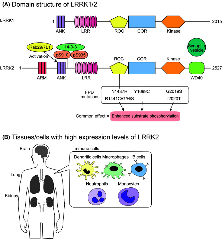
Figure 1. Structure and expression of LRRK2.
(A) The domain structure of LRRK1/2. Both LRRK1 and LRRK2 are composed of an ROC-type GTP-binding domain, a COR domain, a serine/threonine protein kinase domain, and several repeat domains. Eight pathogenic mutations found in LRRK2 are shown below the domain structure of LRRK2, which increase the levels of substrate phosphorylation in vivo. (B) LRRK2 is highly expressed in the brain, lungs, kidneys, and immune cells, including dendritic cells, macrophages, B cells, neutrophils, and monocytes.

