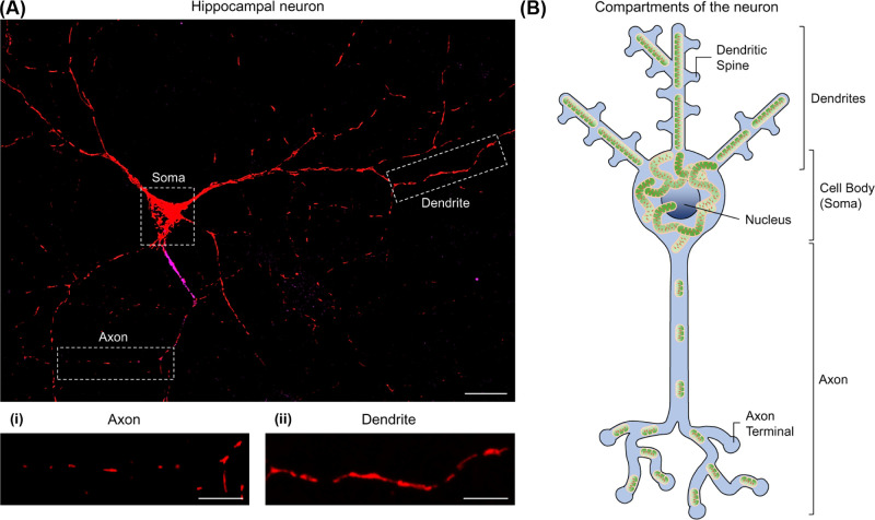
Figure 1. Neuronal mitochondria exhibit compartment-specific morphologies.
(A) Primary rat hippocampal neuron expressing a mitochondrially targeted fluorescent protein (MitoDS-Red). Ankyrin-G staining (magenta) shows the axonal initial segment, used to identify the axon. Highlighted within the boxes are the axonal (i) and dendritic (ii) compartments (enlarged beneath); scale bar: 20 µm, 10 µm in enlargements. (B) Schematic showing the compartments of a neuron, depicting the long axon and multiple dendrites from the cell body (soma), containing the nucleus. Axonal mitochondria are small and sparse, whereas dendritic mitochondria are larger and occupy a greater volume of the process. Mitochondria are densely packed within the soma.

