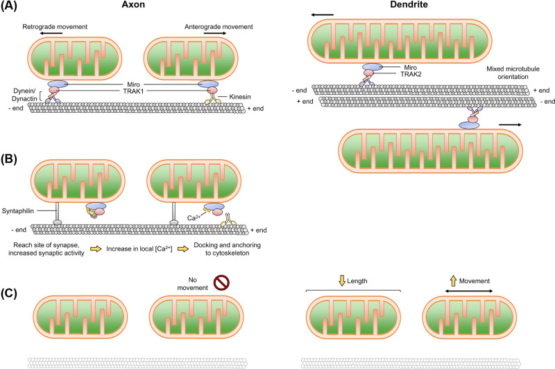
Figure 3. Mitochondrial transport in axons and dendrites.
(A) Mitochondria are transported in an anterograde and retrograde fashion along microtubules via kinesin and dynein/dynactin motor proteins. Microtubules are arranged from minus to plus-ends away from the cell body, whereas microtubules in the dendrite are arranged non-uniformly. The adaptor protein TRAK1 preferentially localises within axons, while TRAK2 preferentially localises to dendrites. (B) Upon an action potential or reaching a site of high energy demand characterised by an increase in [Ca2+]i, a reversible rearrangement of the motor and adaptor complex occurs, causing axonal mitochondria to pause. There are a number of models of how this occurs (i) Miro binds to Ca2+ and induces a conformational change in kinesin, releasing it from the microtubule tract and (ii) Miro binding to Ca2+ causes it to dissociate from kinesin. Syntaphilin also helps to immobilise mitochondria to the microtubule. How/if dendritic mitochondria are stabilised in a similar manner remains to be established. (C) Disruption of the neuronal cytoskeleton (actin and microtubules) stabilises axonal mitochondria, whereas dendritic mitochondria become shorter and more dynamic.

