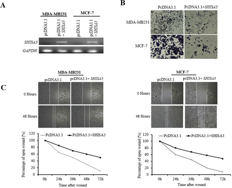
Fig 4. Breast cancer cells proliferation and migration abilities before and after SHISA3 ectopic expression.
A) qPCR of SHISA3 expression in control (pcDNA3.1) and pcDNA3.1-SHISA3 transfected MDA-MB231 and MCF-7 cells. GAPDH was used as internal control for normalized gene expression. B) Cell proliferation assay of MDA-MB231 and MCF-7 cell before and after transfection of SHISA3. C) Wound-healing assay for cell motility of empty vector- or SHISA3-expressing vector transfected MDA-MB231 and MCF-7 cells. Upper: Representative images of wound sealing at 0 h or 48 h after wound scratch. Lower: percentage of wound sealing compared with that of controls at each time point as indicated.

