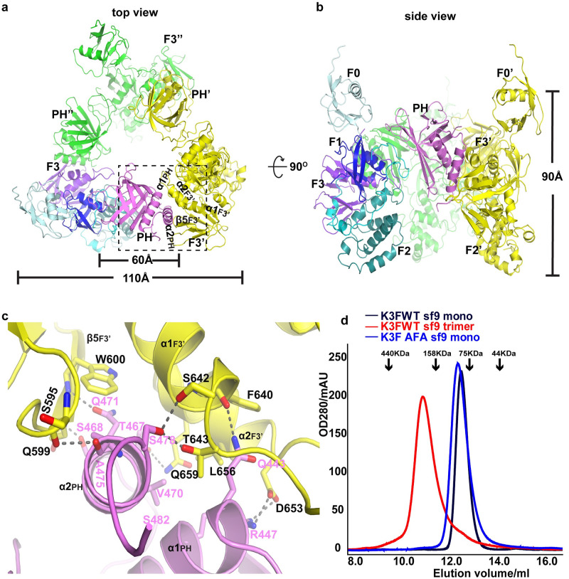
Fig 2. Trimer formation of kindlin-3.
(a) “Top” view of the kindlin-3 homotrimer. One protomer is represented with domain shown in the same color scheme as in Fig 1A, the other 2 protomers are colored yellow and green, respectively. The prime ′ and double prime ″ refer to protomers B and C (same as below) in the trimer, respectively. (b) “Side” view of the kindlin-3 homotrimer. (c) Close-up view of the detailed interactions along the trimer interface between the PH domain of one protomer and the F3 domain of the neighboring protomer. The H-bonds and salt bridges are shown with dashed lines. (d) Analytical gel filtration chromatography profiles of native and mutant kindlin-3 purified from insect cells. K3FWT monomer (black) and trimer (red): the native kindlin-3 expressed in Sf9 insect cells could be prepared as both monomer and trimer; K3FAFA monomer (blue): the kindlin-3 with triple mutations Q471A, A475F, and S478A in trimer interface exhibits monomeric state. Note that molecular weight markers for analytical gel filtration chromatography are indicated by black arrows. AFA, triple mutations Q471A, A475F and S478A; PH, pleckstrin homology; Sf9, Spodoptera frugiperda 9; WT, wild type.

