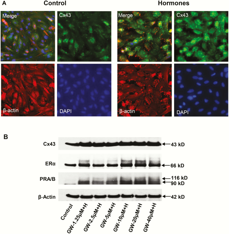
Figure 5.
A, The effects of hormone (“H”) treatment on immunofluorescent connexin 43 (Cx43) (green) staining in endometrial stromal cell (ESC) cultures for 72 hours are shown in the upper right panels. A 2.6 ± 0.5-fold increase in Cx43 pixel count (P < .05, t test, n = 4) was noted. DAPI (4’,6-diamidino-2-phenylindole) nuclear staining (lower right panels), β-actin (lower left panels), and a merged frame (upper left panels) also are presented. B, Western blot results in ESCs treated with different concentrations of peroxisome-proliferator-activated receptor β/δ (PPARβ/δ) agonist (GW) combined with decidualizing hormones (“H”) for Cx43, estrogen receptor α (ERα) and PR-A and PR-B are shown. Standard E2 + P4 + 3′,5′–cyclic adenosine 5′-monophosphate (cAMP) hormone concentrations were kept constant while GW levels were increased. ERα (panel 2) and PR-A and -B (panel 3) both revealed dose-dependent increases by the addition of GW (“GW + H”).

