Figure 2.
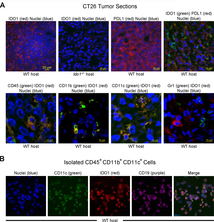
Indoleamine 2,3-dioxygenase 1 (IDO1) expression is localized to infiltrating immune cells within CT26 tumors. (A) Confocal images of CT26 tumor sections. (top row, left to right) Immunofluoresence staining of sections from wild-type (WT) and Ido1-/- mice for IDO1 (Cy3, red), and from WT mice for programmed death-ligand 1 (PDL1) (Cy3, red) and the combination of IDO1 (fluorescein isothiocyanate (FITC), green) and PDL1 (Cy3, red). Nuclei were stained on all sections (DAPI, blue). (bottom row, left to right) Staining of CT26 tumor sections from WT mice for combinations of IDO1 (Cy3, red) with CD45, CD11b, Gr1 and CD11c (FITC, green). Nuclei were stained on all sections (DAPI, blue). (B) Confocal images of a field of FACS-isolated CD45+, CD11b+, CD11c+ cells from a CT26 tumor in a WT host. (left to right) Immunofluoresence staining for nuclei (DAPI, blue), CD11c (FITC, green), IDO1 (Cy3, red), CD19 (Cy5, purple) and the composite image.

