. 2020 Apr 21;296(1):76–84. doi: 10.1148/radiol.2020190646

Figure 2b:

Images show representative peripheral zone lesions (arrow). (a) Prostate Imaging Reporting and Data System (PI-RADS) score 2. T2-weighted MRI scan shows triangular lesion with low T2 signal intensity in right midgland. (b–d) PI-RADS score 3. T2-weighted MRI scan (b), diffusion-weighted MRI scan obtained with high b value (c), and apparent diffusion coefficient (ADC) map (d) show focal lesion in left apex. Lesion has mildly low signal intensity on ADC map and mildly high signal intensity on diffusion-weighted image. No enhancement was seen on dynamic contrast material–enhanced image (not shown). (e–g) PI-RADS score 4. T2-weighted MRI scan (e), diffusion-weighted MRI scan obtained with high b value (f), and ADC map (g) show 1.0-cm focal lesion in right midgland. Lesion has markedly low signal intensity on ADC map and markedly high signal intensity on diffusion-weighted MRI scan. (h) PI-RADS score 5. T2-weighted MRI scan shows large left midgland lesion associated with extraprostatic extension.

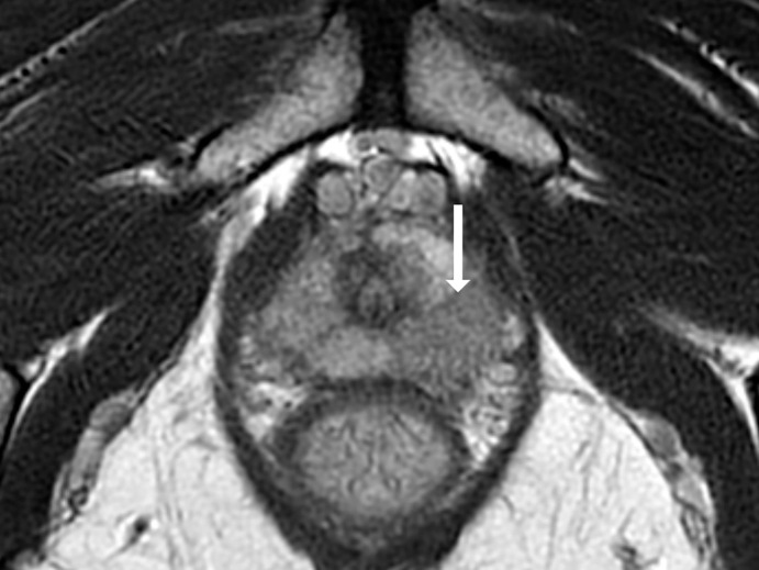
Images show representative peripheral zone lesions (arrow). (a) Prostate Imaging Reporting and Data System (PI-RADS) score 2. T2-weighted MRI scan shows triangular lesion with low T2 signal intensity in right midgland. (b–d) PI-RADS score 3. T2-weighted MRI scan (b), diffusion-weighted MRI scan obtained with high b value (c), and apparent diffusion coefficient (ADC) map (d) show focal lesion in left apex. Lesion has mildly low signal intensity on ADC map and mildly high signal intensity on diffusion-weighted image. No enhancement was seen on dynamic contrast material–enhanced image (not shown). (e–g) PI-RADS score 4. T2-weighted MRI scan (e), diffusion-weighted MRI scan obtained with high b value (f), and ADC map (g) show 1.0-cm focal lesion in right midgland. Lesion has markedly low signal intensity on ADC map and markedly high signal intensity on diffusion-weighted MRI scan. (h) PI-RADS score 5. T2-weighted MRI scan shows large left midgland lesion associated with extraprostatic extension.