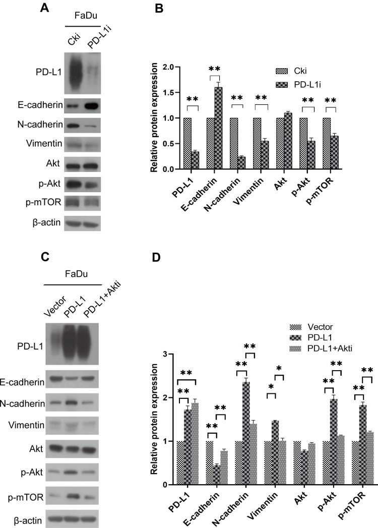
Figure 4.
PD-L1 promoted EMT through the Akt-mTOR signaling pathway. (A) FaDu cells were transfected with PD-L1 siRNA or control siRNA, expression of PD-L1, E-cadherin, N-cadherin, Vimentin, Akt, p-Akt and p-mTOR was analyzed by Western blot. β-actin was used as an inner control. (B) Bands of Western blot were analyzed by Image J software. Results were obtained from the ratio of target band to β-actin. The data are presented as means ± standard deviation from three independent experiments. **P<0.01. (C) PD-L1-overexpressed or control FaDu cells were treated with Akt-inhibitor. The indicated protein levels were assayed by Western blot. β-actin was used as an inner control. (D) Bands of Western blot were analyzed by Image J software. Results were obtained from the ratio of target band to β-actin. The data are presented as means ± standard deviation from three independent experiments. *P<0.05, **P<0.01.
Abbreviations: PD-L1, programmed death-ligand 1; EMT, epithelial–mesenchymal transition; siRNA, small interfering RNA; Cki, control siRNA.

