
Figure 3.
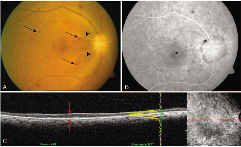
Fundus photography, fluorescein angiography and optical coherence tomography (OCT) of case 2. (A) Fundus photography shows slight blurring and an atrophic optic disc with dot and blot retinal hemorrhages (arrows) in the posterior pole. The retinal arteries are attenuated, and vessels show sheathing (arrowheads). (B) Fluorescein angiography findings are consistent with fundus photography. Macular and peripapillary areas show uneven choroidal filling (asterisks). (C) OCT shows thin atrophic neurosensory retina with indistinct retinal layers (red arrows).