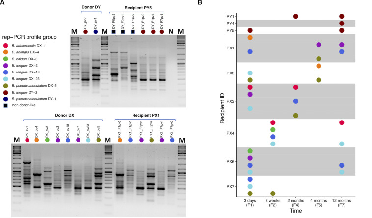
FIGURE 2.
Rep-PCR typing of bifidobacteria isolated from fecal samples of FMT donors and recipients. (A) Representative gel pictures presenting rep-PCR fingerprint profiles of bifidobacterial isolates from donors DY and DX and their respective recipients PY5 and PX1 at different time points: PY5 has unique profiles pre-FMT, but also DY-like profiles post-FMT (upper gel). PX1 has DX-like profiles post-FMT (lower gel). (B) A visual summary of donor-like rep-PCR fingerprint profiles observed among each recipient’s isolates during the 1-year follow-up after FMT. Each colored dot refers to a distinct type of rep-PCR fingerprint profile observed among the donor isolates. A profile was indexed based on the first isolate to have that profile. For instance, purple dot representing profile B. longum DX-2 was indexed according to the donor isolate B. longum DX_pv2 in which the profile was encountered for the first time. If such profile was observed in a recipient isolate, the isolate was considered donor-like and received the same color. For instance, recipient isolate B. longum PX1_F7pv1 was observed to have B. longum DX-2 profile and received the purple color. Thus, all the donor and donor-like isolates with the same profile were considered representatives of a same rep-PCR profile group and putatively being of the same origin. All the profiles observed among recipient isolates but not in donor isolates were referred collectively with a black square (A). PY1 and PY4-5, FMT recipients of DY; PX1-4 and PX6-7, FMT recipients of DX. The isolate code includes reference to the sample from which it was isolated, see Supplementary Table 2. M, GeneRulerTM DNA Ladder Mix; N, negative PCR control.

