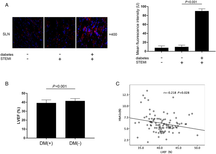
FIGURE 6.

(A) Captured images of SLN fluorescence inmmunohistochemistry staining of human cardiac tissue sampled from patients diagnosed as degenerative valvular disease (without diabetes and coronary arterial disease, n = 6), patients diagnosed as ST‐segment elevation myocardial infarction (STEMI) without diabetes (n = 6), and patients diagnosed as STEMI complicated with diabetes (n = 6) who were undergoing cardiac surgeries. (B) Comparisons of left ventricular ejection fraction (LVEF%) in diabetic and non‐diabetic STEMI patients. (C) The correlation analysis between HbA1c% and LVEF% in STEMI patients.
