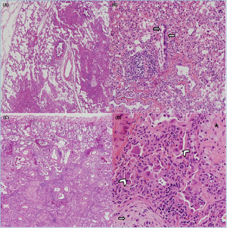
Fig. 3.

Pathologic findings of lung biopsy in a humidifier disinfectant-associated interstitial lung disease case. (A) In the early phase, centrilobular distribution of interstitial thickening and fibrosis was observed (H&E, ×40). (B) Disrupted bronchioles infiltrated by lymphocytes and histiocytes were observed in the early phase (H&E, ×200). (C) In the advanced phase, interstitial thickening with fibrosis was observed with centrilobular distribution and relative sparing of the subpleural parenchyma (H&E, ×40). (D) Extensive interstitial fibroblast proliferation was seen in the pale myxoid stroma (arrows) and collapsed alveolar spaces were lined by activated pneumocytes (arrowhead). Intra-alveolar collection of foamy histiocytes (asterisk) was observed (H&E, ×200).
