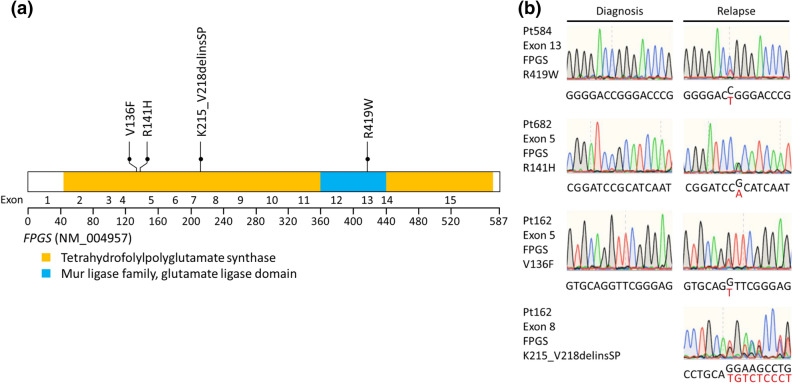
Figure 2.
FPGS mutations in relapsed pediatric ALL. (a) Schematic representation of the structure of the FPGS protein. Tetrahydrofolylpolyglutamate synthase, Mur ligase family, and glutamate ligase domains are indicated. FPGS mutations identified in relapsed pediatric samples are shown. Filled circles represent heterozygous mutations. (b) DNA sequencing chromatograms of paired diagnosis and relapse genomic ALL DNA samples showing representative examples of relapse-specific heterozygous FPGS mutations, with the mutant allele sequence highlighted in red.

