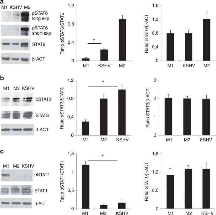
Fig. 3. KSHV infection activates STAT3 and STAT6 in macrophages.
a STAT6, b STAT3 and c STAT1 activation in M1, M2 and KSHV-infected macrophages was evaluated by western blot analysis. β-actin (β-ACT) was used as loading control. A representative experiment out of three is shown. Histograms represent the mean plus S.D. of the densitometric analysis of the ratio of pSTAT/STAT and STAT/ β-ACT. For pSTAT6 the short exposure was used for densitometric analysis. *p-value < 0.05 was calculated between KSHV-infected macrophages and M1 polarised cells.

