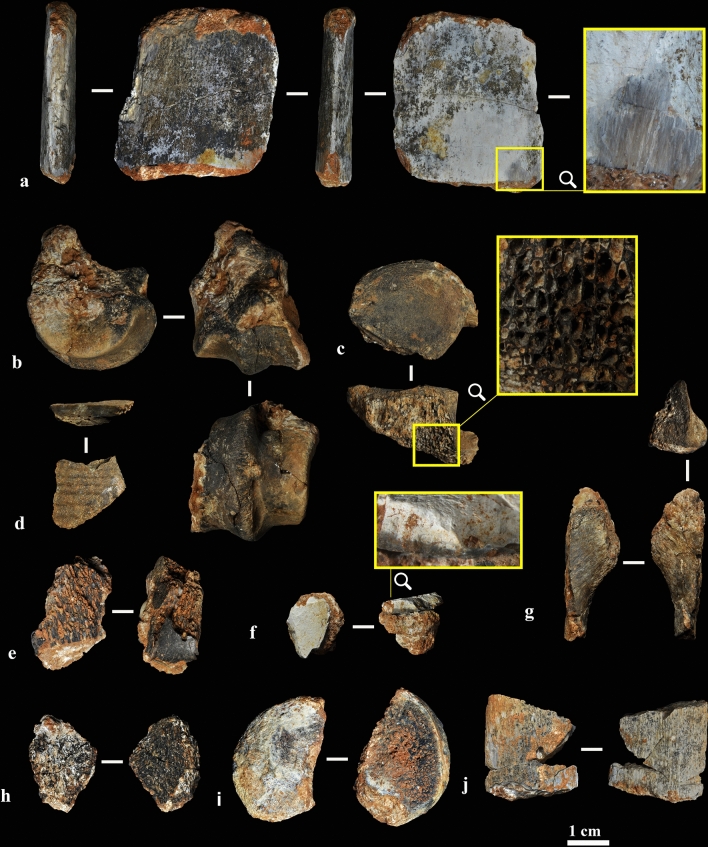
Figure 2.
Burnt bones. (a) Fully calcined rib, blue-grey and white in colour (sample ID #13). (b) Distal condyle of a deer metapodial, fully carbonized (sample ID #3). (c) Fully carbonized vertebral body (sample ID #30). (d) Partially carbonized tortoise bone plate (sample ID #6). (e) Fully carbonized spongy fragment (sample ID #14). (f) Fully calcined flat bone (sample ID #15). (g) Fully carbonized flat bone (sample ID #2). (h) Fully carbonized spongy fragment (sample ID #20). (i) Fully carbonized fragmented epiphysis (sample ID #4). (j) Fully calcined rib (sample ID #27).

