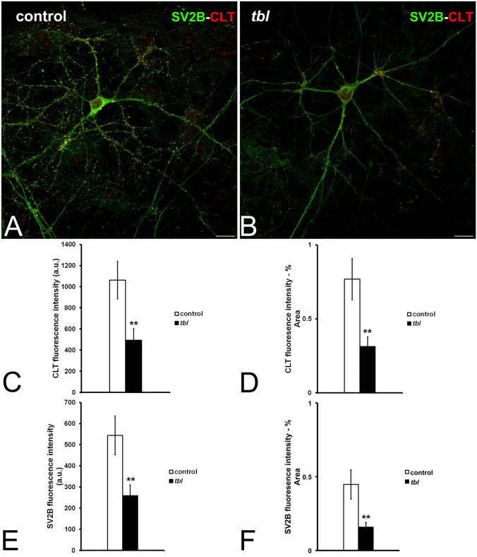
Figure 4.
Laser confocal microphotographs illustrative of SV2B (green) and CLT (red) immunoreactivities in control (A) and tbl (B) hippocampal cultures. (C–F), graphs showing the fluorescence intensities of CLT (C, D) and of synaptic vesicles identified by SV2A immunoreactivity (E,F). In both absolute and % relative values, fluorescence intensity was higher in control than in tbl cultures (C, **p = 0.004; D, **p = 0.002; E, **p = 0.004; F, **p = 0.003). Bars = 30 µm. a.u., arbitrary units.

