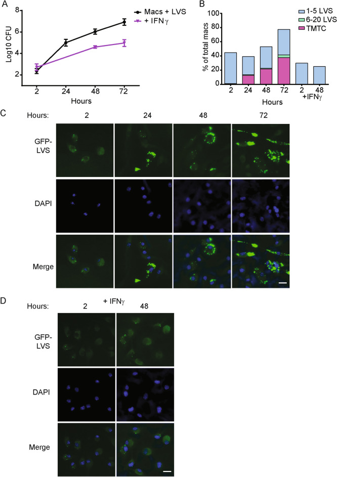
Figure 1.
In vitro intracellular growth and intercellular spread of LVS in mouse BMM. Murine BMM were cultured without or with 50 ng/ml recombinant mouse IFN-γ for 24 h, as indicated, and then infected with GFP-LVS at an MOI of 50:1 (bacteria to macrophage ratio). (A) At the indicated time points after infection, macrophages were washed, lysed, and plated to evaluate the recovery of intracellular bacteria. Values shown are the mean numbers of CFU/ml of viable bacteria ± SD of triplicate samples. (B) GFP-LVS-infected macrophages were scored by visual inspection for numbers of bacteria in each macrophage. The level of infection was categorized into four groups: macrophages containing no bacteria, 1–5 bacteria, 6–20 bacteria, or > 21 bacteria as too many to count (TMTC). At least 100 macrophages from two identical replicates were scored for each condition. (C, D) Representative images of BMM infected with GFP-LVS ± IFN-γ at indicated time points after infection. Scale bar = 50 µM. Results shown are from one representative experiment of three independent experiments of similar design and outcome.

