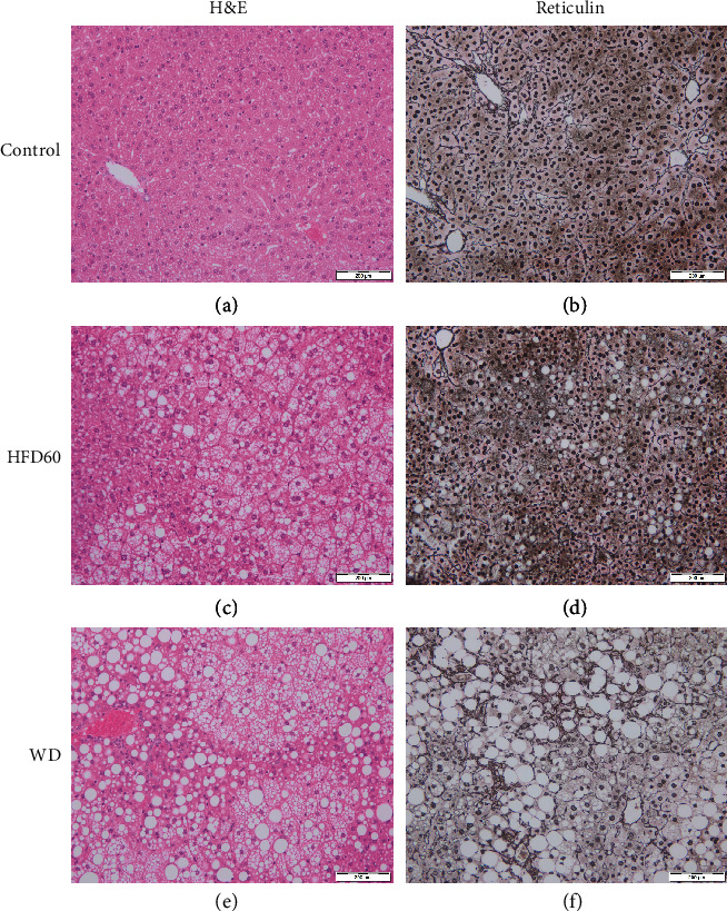
Figure 3.

H&E- and reticulin-stained liver sections. Representative histological images of (a, b) control, (c, d) HFD60, and (e, f) WD mice at 12 weeks. Images show severe steatosis, especially in HFD60 livers, and fibrosis after WD treatment. Magnification 200x, scale bars 200 μm.
