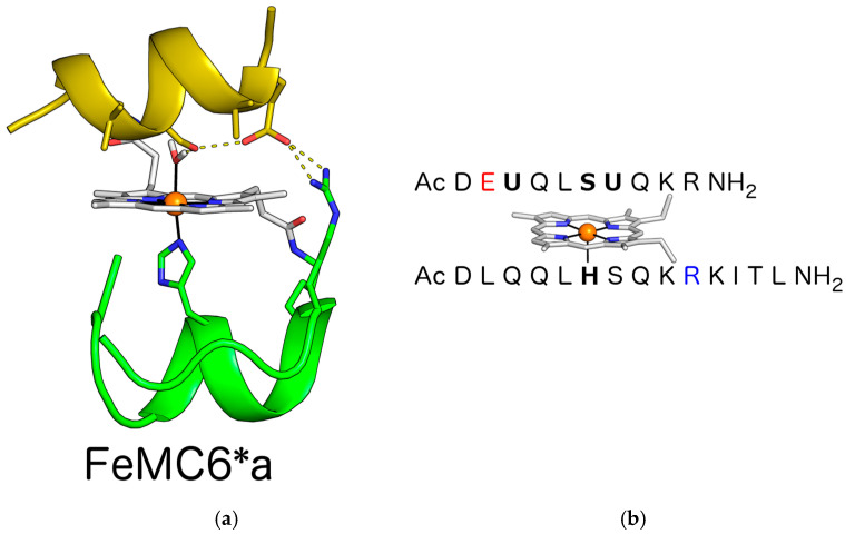
Figure 1.
(a) FeMC6*a designed model. Key functional residues and the deuteroporphyrin IX are highlighted as sticks. Iron is represented as an orange sphere. Aib residues face the porphyrin ring at the distal site. The designed inter-chain ion pair interaction is depicted, together with the hydrogen bond network presumably involved in hydrogen peroxide activation. (b) FeMC6*a peptide sequences. Proximal His and distal axial residues are indicated in bold. The Glu and Arg residues involved in the inter-chain ion pair interaction are depicted in red and blue, respectively.

