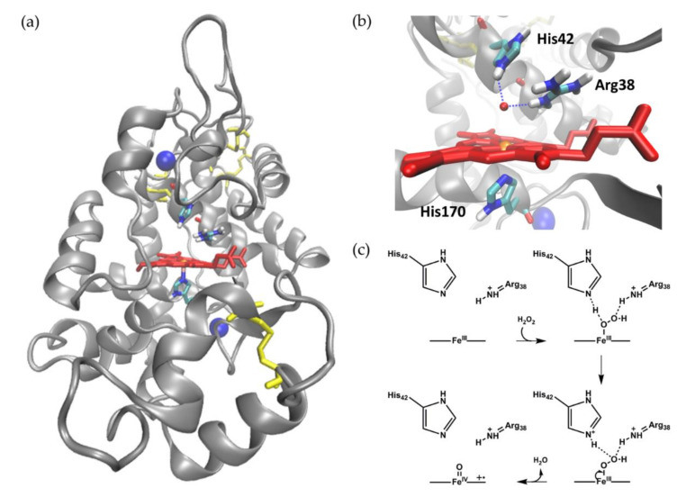
Figure 1.
Crystal structure of HRP and mechanism for hydrogen peroxide reduction. (a) Overall globular structure of HRP C1A. The polypeptide chain is shown as the grey cartoon, the heme cofactor, disulfide bonds, and selected amino acids as red, yellow, and multi-colored sticks, respectively. The two calcium ions are shown as blue spheres. (b) Active site of HRP C1A with the heme cofactor depicted as red sticks with the iron center as orange sphere. The essential amino acids Arg38, His42, and His170 are shown as sticks colored by element. Structures were visualized using PDB 1ATJ [33] and VMD 1.9.3 [37]. From Neumann, 2019 [38]. (c) Proposed mechanism for hydrogen peroxide reduction by heme peroxidases and concomitant Compound I formation. Adapted with permission from Rodríguez-López et al., 2001 [34]. Copyright (2001) American Chemical Society.

