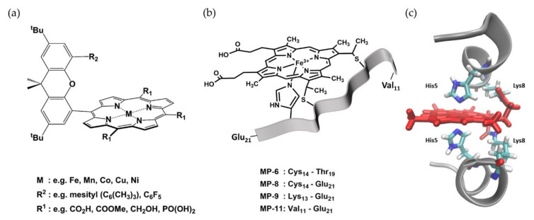
Figure 3.
Structures of selected peroxidase mimics. (a) Chemical structure of a Hangman porphyrin with a xanthene linker. Various combinations of meso-substituents (R1), metal centers (M), and hanging groups (R2) have been reported. Examples for each functionality are given below. Taken from [38]. (b) Illustration of microperoxidases with the heme in black lines and the polypeptide chain as a grey ribbon. The respective polypeptide segments of the different microperoxidases are noted below. (c) Crystal structure of Co-mimochrome IV with polypeptide chains as grey cartoon, the heme cofactor, and selected amino acids as red and multi-colored sticks, respectively. PDB 1PYZ [50] visualized with VMD 1.9.3. [38].

