
Fig. 2.
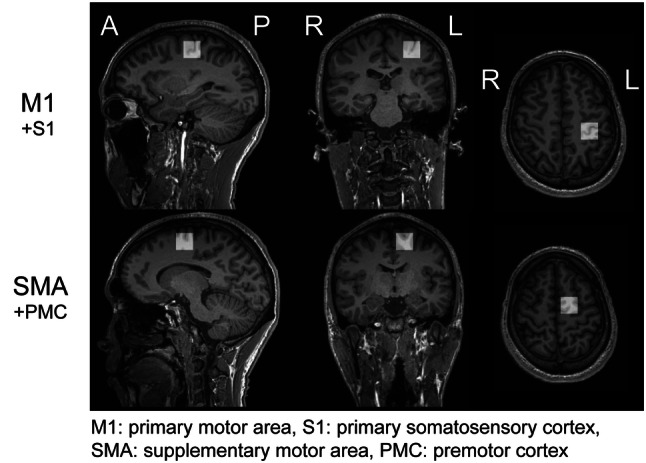
ROIs for 1H-MRS data acquisition. ROIs were placed in the left M1 and left SMA with a voxel size of 20 × 20 × 20 mm3. The M1 ROI was placed in the left precentral sulcus, and the midpoint of the SMA ROI was set as the upper part of the Brodmann area 6. The ROIs in the M1 and SMA areas included slightly the primary somatosensory cortex (S1) and premotor cortex (PMC), respectively. For both ROIs, the inclusion of bone and cerebrospinal fluid was avoided