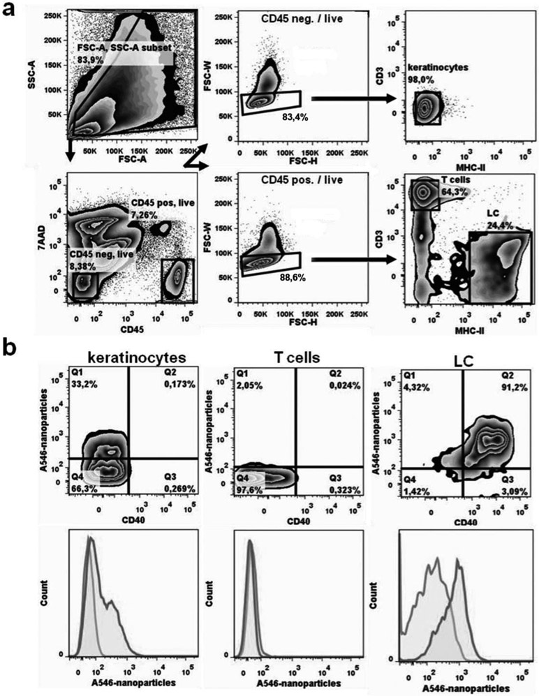
Figure 1. In vitro capture of pDNA/PEIm nanoparticles by LCs.
After skin preparation epidermal cell suspensions were isolated and incubated with Alexa-546-labeled nanoparticles (A546-nanoparticles). a) Representative FACS gating strategy. Arrows indicate the sequence of steps. First, cells are discriminated from debris on the basis of forward (FSC-A) and side scatter (SSC-A) characteristics (top left panel). Second, dead cells are excluded from further analysis by their 7AAD positivity (lower left panel). Third, within viable CD45-negative cells (i.e., keratinocytes; upper middle panel) and viable CD45+ cells (lower middle panel) doublets are identified by their FSC-W and FSC-H properties and are omitted from further analysis. Doublets are the cells outside the gated area; about 20%. Single cells (about 80%) are within the gate. Fourth, epidermal cells are defined as MHC-II-/CD3- keratinocytes (upper right panel) and MHC- II+/CD3- Langerhans Cells and MHC-II-/CD3+ dendritic epidermal T cells / DETC (lower right panel). b) Representative FACS contour plots (upper row) and histogram overlays (lower row) of keratinocytes, dendritic epidermal T cells and CD40+ Langerhans cells show the uptake of nanoparticles. show the uptake of Alex-546-conjugated nanoparticles (dark grey lines) compared to control cultures without nanoparticles (light grey lines). Representative FACS plots are shown of 4 individually analysed mice per group.

