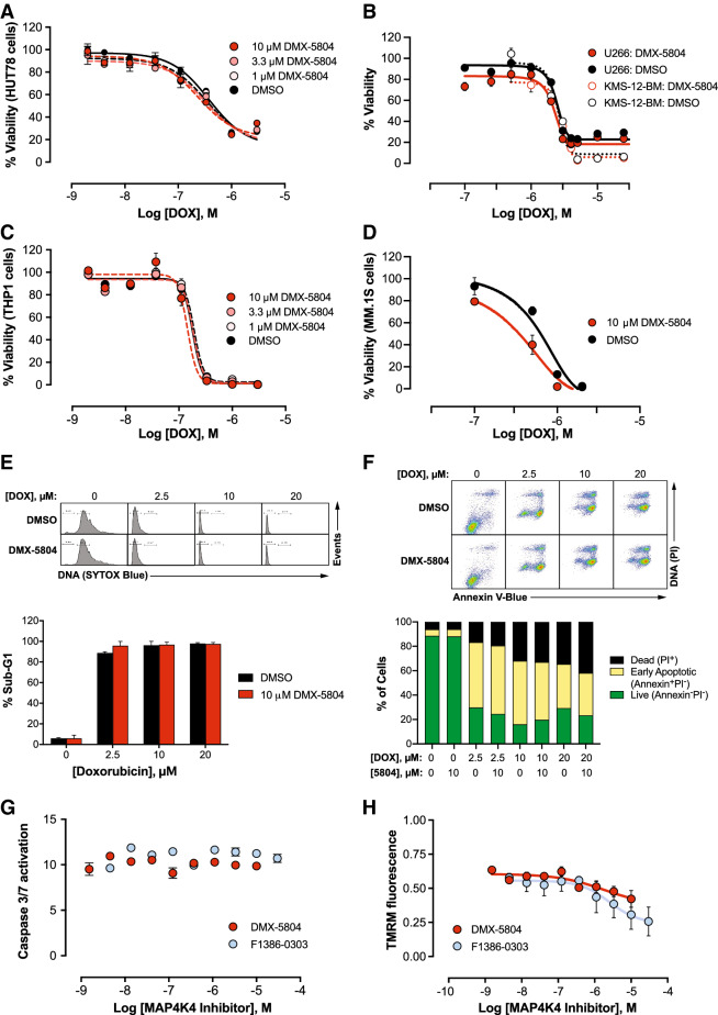
Figure 3.
Inhibitors of MAP4K4 do not impair cancer cell killing by DOX. (A–D) Viability. Human tumor cells from the five indicated lines were treated with DOX and DMX-5804 at the concentrations shown and were assayed at 24 h (CellTiter Glo). Data are the mean ± SEM of ≥ 5 replicates and are representative of 2 independent dose–response experiments, excepting KMS-12-BM (5 replicates, 1 study). (E) Hypodiploid (sub-G1) DNA fragmentation. U266 cells were treated with 0–20 μM DOX for 24 h ± 10 μM DMX-5804, and DNA content was analysed by flow cytometry. Above, representative DNA histograms. Below, results are the mean ± SEM of single determinations in 2–3 independent experiments. (F) Annexin V. U266 cells were treated as in panel E then were analysed by flow cytometry for annexin V-Blue and PI fluorescence. Above, Representative scattergrams. Below, Data are single determinations, representative of two independent experiments. (G) Activation of caspase-3/7 (cf. Fig. 1E). THP1 cells were treated with 1 μM DOX for 24 h ± F1386-0303 at the concentrations shown. Data are the mean ± SEM of 4 replicates in each of 3 independent dose–response experiments, shown relative to the vehicle-treated control. (H) Loss of mitochondrial membrane potential. (cf. Fig. 1H). THP1 cells were treated with 1 μM DOX for 16 h ± F1386-0303 at the concentrations shown. TMRM fluorescence is shown as the mean ± SEM of 4 determinations in each of 3 independent dose–response experiments, relative to the vehicle-treated control.

