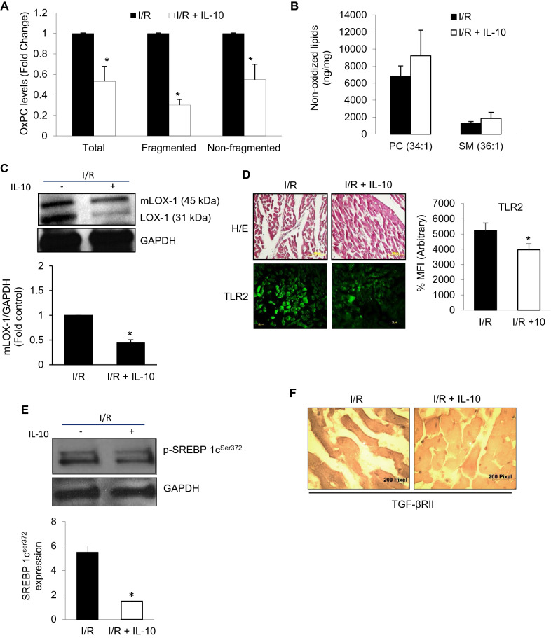
Figure 5.
IL-10 modulates TLR2 innate signaling and OxPCs-mediated changes in global ischemia–reperfusion (I/R) injury. Hearts were subjected to global I/R with and without IL-10 as described in the methods. For IL-10 treatment, 10 ng/ml of IL-10 was added for 40 min during the reperfusion phase. (A) Total, fragmented and non-fragmented OxPCs were measured using mass-spectrometer for each fraction and their levels in I/R hearts were normalized compared with those in the presence of IL-10. (B) Non-oxidized lipids (PC Phosphatidylcholine and SM Sphingomyelin). (C) Western blotting was used to determine protein expression of mLOX1 and LOX1 post-I/R. (D) Expression of TLR2 was detected in the myocardium using immunofluorescence. Percentage of mean fluorescence intensity (% MFI) was calculated from three different heart sections and counted in 10 different fields after TLR2 staining. Upper panel is showing for Hematoxylin and Eosin (H/E) staining. (E) Western blots were probed with antibody for SREBP 1cser372 . GAPDH was used as a loading control. Histogram (E, lower panel) is representative of 3 independent hearts probed for SREBP1cser372. (F) TGF-β receptor II (TGF-βRII) expression was also studied in tissue sections using ABC peroxidase staining. Sections were probed with specific TGF-βRII antibody followed by diaminobezidine (DAB) staining.

