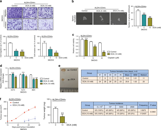
Fig. 5. DCA hinders ovarian CSC properties in vitro and in vivo.
a Transwell migration/invasion (scale bar, 100 μΜ) and b sphere formation assays (scale bar, 50 μΜ) of ALDH+CD44+ SKOV3 cells after treatment with vehicle and DCA (5 and 10 mM) for 48 h. c Relative cell viability of vehicle or 10 mM DCA-treated ALDH+CD44+ SKOV3 cells after treatment with or without 20 μM cisplatin for 48 h with the XTT assay. d qPCR analysis of mRNA expression of relative stemness genes, OCT4, KLF4 and BMI1, of ALDH+CD44+ SKOV3 cells after treatment with DCA (5 and 10 mM). Following injection of 3 × 10⁵ ALDH+CD44+ SKOV3 cells pre-treated with control (n = 5) DCA (n = 5) into the right/left flanks of mice. e (Left) Representative images of xenograft tumours and (right) time of tumour formation (days) in the xenograft model were recorded. f (Left) Tumour size curves and (middle) weights of xenografts derived from control (n = 5) and DCA (n = 5)-pre-treated ALDH+CD44+ SKOV3 cells. (Right) The tumour initiation rate was recorded based on the in vivo limiting dilution assay (*p < 0.05, **p < 0.01, results represent means ± SD of three independent experiments; n.s., not significant).

