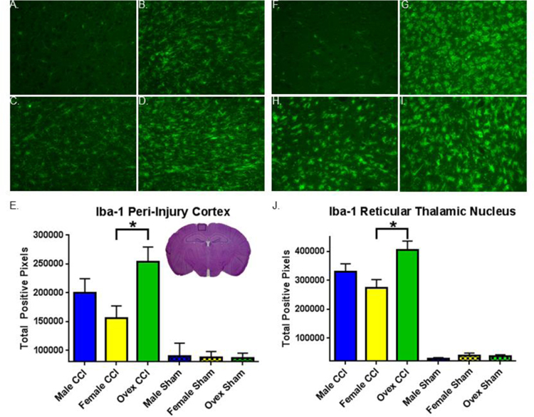
Figure 4. Iba-1 immunohistochemistry varies by group.
A.–D. Representative images of Iba-1 immunofluorescent staining in peri-injury cortex (CTX). A. Sham CTX. B. Male CTX. C. Female CTX. D. Ovariectomized female (ovex) CTX. E. Quantification of total positive Iba-1 pixels / image in the peri-injury cortex. Bars indicate the group mean. Error bars indicate standard error of the mean. Injury > sham, p< 0.0001. Ovex CCI > female CCI, *p = 0.01. F.–I. Representative images of Iba-1 immunofluorescent staining in the reticular thalamic nucleus (RTN). F. Sham RTN. G. Male RTN. H. Female RTN. I. Ovex RTN. J. Quantification of total positive Iba-1 pixels / image in the RTN. Injury > sham, p< 0.0001. Ovex CCI > female CCI, *p = 0.0002.

