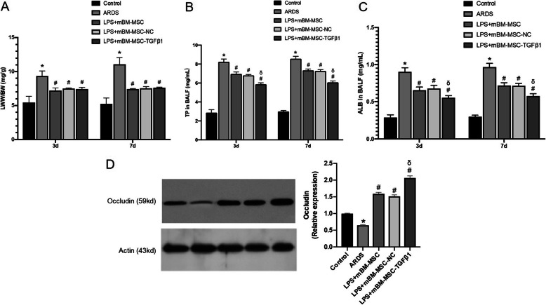
Fig. 7.
The effect of mMSCs overexpressing TGFβ1 on the permeability of LPS-induced lungs in mice. a Lung oedema was analysed by LWW/BW (n = 3, *p < 0.05 compared with the control group; #p < 0.05 compared with the ARDS group). b Total protein and c albumin concentrations in bronchoalveolar lavage fluid were analysed by a mouse-specific ELISA kit to evaluate the permeability of the lung (n = 3, *p < 0.05 compared with the control group; #p < 0.05 compared with the ARDS group; δp < 0.05 compared with the LPS + mBM-MSC-NC group). d Expression of the occludin protein in the lungs of all the experimental groups at 3 and 7 days after mMSCs administration was evaluated by western blot analysis (n = 3, *p < 0.05 compared with the control group; #p < 0.05 compared with the ARDS group; δp < 0.05 compared with the LPS + mBM-MSC-NC group). ALB, albumin; ARDS, acute respiratory distress syndrome; ELISA, enzyme-linked immunosorbent assay; LWW/BW, lung wet weight/body weight; mMSCs, mouse mesenchymal stem cells; NC, normal control; TP, total protein

