Fig. 4.
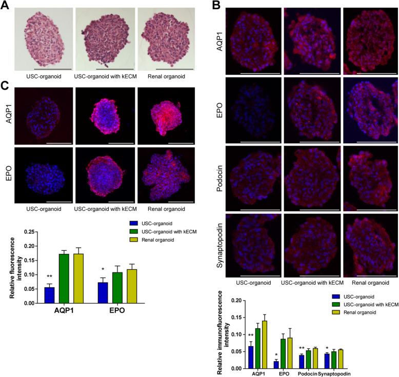
Histology of 3D organoids (USC-organoids, USC-organoids with kECM, and renal organoids). a H.E. staining. The cell nucleus was blue stained, and cytoplasm was red stained. b Immunofluorescence staining for specific proximal tubule AQP1, kidney endocrine product EPO, and kidney glomerular markers Podocin and Synaptopodin. AQP1, EPO, and Podocin and Synaptopodin were labeled as red and DAPI as blue. The fluorescence intensity was quantified. c Whole mount staining for AQP1 and EPO. Red fluorescence indicated AQP1 and EPO and DAPI as blue. The whole mount staining provided 3D vision. The fluorescence intensity was quantified (scale bar 200 μm)

