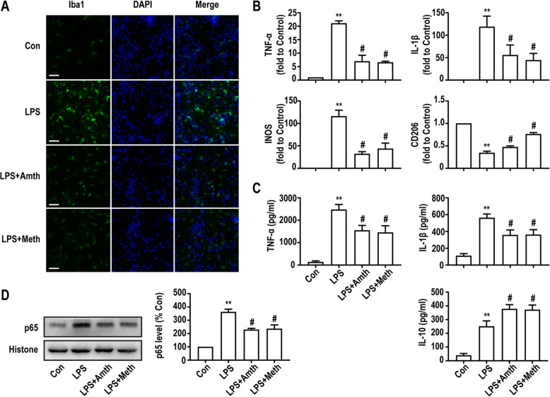
Fig. 6.
An agonist of H2R or H3R inhibits LPS-induced microglia activation and associated inflammatory response. a Primary microglia were stained with Iba1 antibody as indicated. Blue staining represents DAPI. Scale bar = 50 μm. b q-PCR analysis of the relative expression of TNF-α, IL-1β, INOS, and CD206 mRNA. Each value was expressed relative to that in the control group, which was set to 1. c Concentration of TNF-α, IL-1β, and IL-10 measured by ELISA. d The expression levels of NF-κb were detected in primary cultured microglia by Western blotting using specific antibodies. Expression of NF-κb was quantified and normalized to histone levels. Each value was expressed relative to that in the control group, which was set to 100. **P < 0.01 versus the control group. #P < 0.05 versus the LPS group (n = 3). The data are presented as the mean ± SEM

