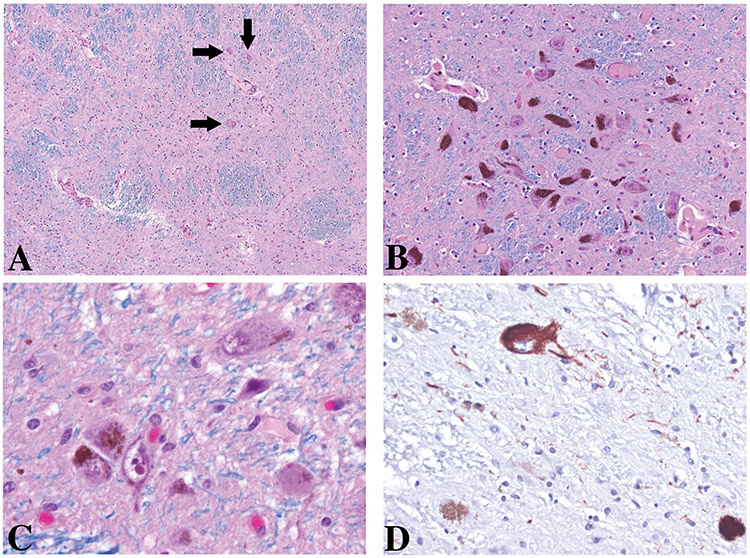
FIGURE: Substantia Nigra.
Uneven loss of pigmented neurons: subtotal loss within the lateral third at the level of the decussation of the superior cerebellar peduncle of the pars compacta (A), rare residual neurons are present (arrows). Relative spared areas within the intermediate third (B) and lateral third (C and D) of the pars compacta at the level of the red nucleus. Among the residual pigmented neurons a few are labeled with AT8 antibodies (D). No Lewy body-containing neurons are present throughout the sections.
A, B, and C: Luxol-Hematoxylin and Eosin; D, tau antibodies (AT8) Original magnification 100X, 200X, 630X, and 400X.

