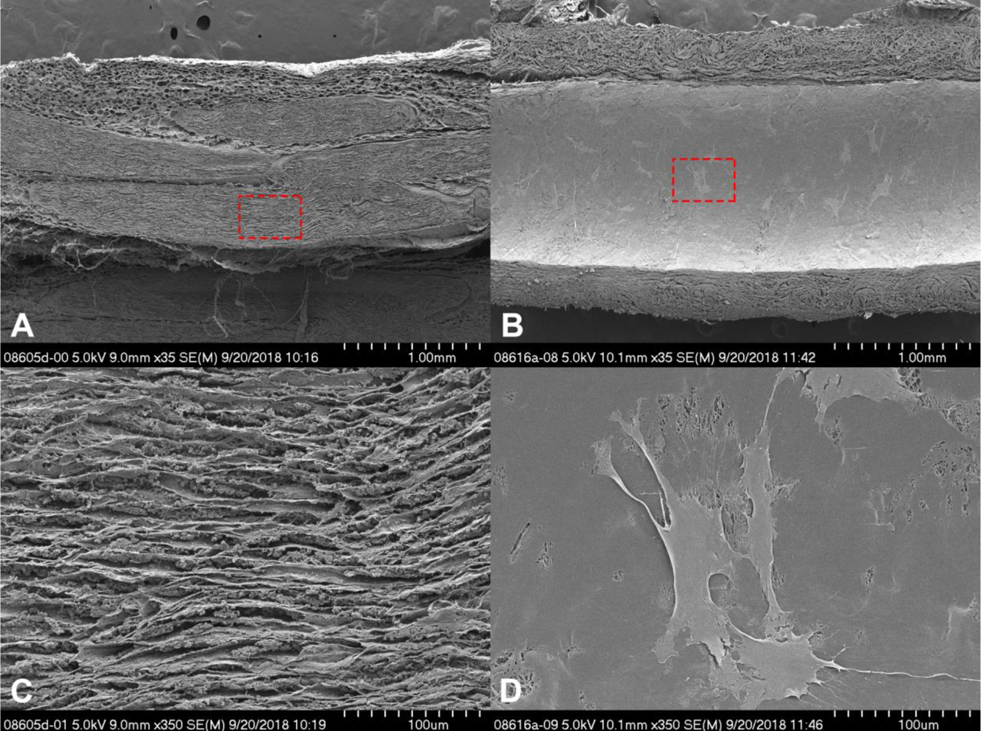
Figure 6.

Cross-sectional scanning electron microscopy images of the Avance® Nerve Graft (A and C) and the NeuraGen® Nerve Guide (B and D) after 12 h of dynamic seeding with human MSCs. Both nerve substitutes were cut longitudinally. The cross-section of the Avance® Nerve Graft shows aligned fascicles without the presence of any cells. The cross-section of the NeuraGen® Nerve Guide demonstrates the smooth inner surface of the hollow conduit, with MSCs spread out among the entire length of the nerve guide.
