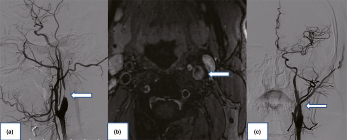
FIGURE 1.

(a) Digital subtraction angiography showing the significant stenosis of the left internal carotid artery (arrow). (b) High‐resolution magnetic resonance imaging showed ruptured fibrous caps, large lipid core, intraplaque hemorrhage, and ulcer (arrow). (c) Completion angiogram after angioplasty and stenting of the left internal carotid artery showing the excellent restoration of flow (arrow)
